Research
Seeing Brain Cells Without Cutting
When people think of MRI, they picture images of the whole brain — regions, tracts, maybe tumors. But what if we could use MRI to look at individual brain cells — neurons and glia — in living humans?
That’s what we set out to do in our latest study, “Resolving Cellular Morphology in the Human Brain with Multiparametric Diffusion MR Spectroscopy”.
Why We Did This
Many neurological diseases — Alzheimer’s, Parkinson’s, multiple sclerosis — involve changes in cell shape and size long before tissue loss is visible on standard MRI. We wanted to find a way to measure those microscopic changes noninvasively.
Following the Metabolites
Standard diffusion MRI tracks water, but water moves everywhere.
We instead followed metabolites — small molecules that live mainly inside specific cell types:
- N-acetylaspartate (NAA): neurons
- myo-inositol (mI) and choline: glial cells
- creatine: both
By measuring how these metabolites move, we can infer cell size, shape, and crowding inside neurons or glia.
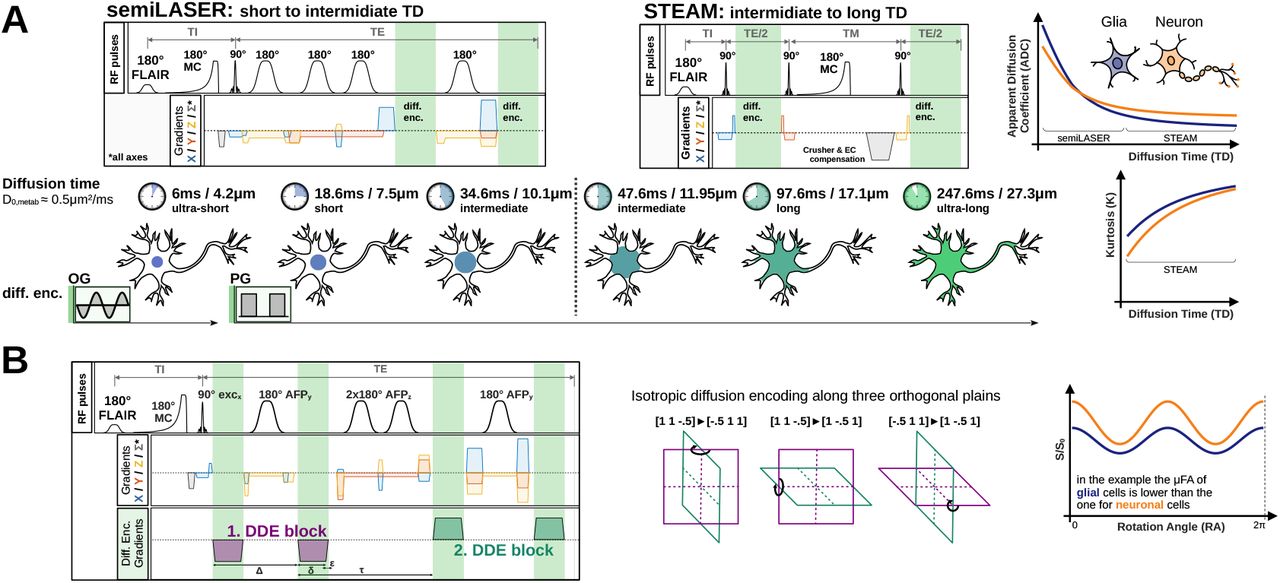
Combining Two Diffusion Techniques
We combined two complementary MRI measurements:
- Diffusion-time experiments — measuring how far molecules move over different timescales (6–250 ms).
- Double-diffusion encoding (DDE) — changing the angle between diffusion gradients to tell whether diffusion occurs in cylindrical (neurites) or spherical (soma) environments.
Together, these form a multiparametric dataset that gives a much clearer picture of cellular structure.
What We Found
Scanning healthy volunteers on strong 3T MRI scanners, we could:
- Observe how metabolite diffusion slowed and became more “non-random” at longer diffusion times — a sign of molecules hitting cell boundaries.
- Estimate realistic cell dimensions:
- Neuronal somas ≈ 6–8 µm
- Glial somas ≈ 9 µm
- Neurite diameters ≈ 1 µm
- Show that combining both diffusion methods produced stable, physically realistic models — avoiding the ambiguities of older approaches.
- Simultaneously measure water diffusion, revealing exchange times between compartments of 13–17 ms.
Why It Matters
This technique turns MRI into a “virtual microscope”.
It could help detect or track diseases that alter cellular structure — from Alzheimer’s to multiple sclerosis — long before tissue damage becomes visible.
And while our current setup used powerful research scanners, a streamlined clinical protocol could fit into under an hour, making it feasible for patient studies.
Looking Ahead
We’re excited by what this means: MRI that doesn’t just show where something happens in the brain, but how cells themselves are shaped and connected.
It’s a step toward noninvasive, cell-specific imaging — and perhaps one day, toward diagnosing and monitoring neurological diseases at the cellular level.